Herzlich Willkommen im
DEUTSCHEN CYBERKNIFE ZENTRUM
Präzise Strahlung auf den Punkt gebracht

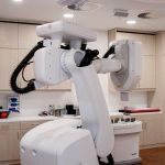
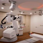
Fortschrittlichste Technik
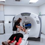
Die CyberKnife®-Roboter gehören zur medizinischen Hochtechnologie und ermöglichen eine schonende und präzise Behandlung.
Fachleute mit viel Erfahrung
Sie haben die besten Spezialisten verdient. Deshalb arbeiten wir mit vielen namenhaften Ärzten und Fachleuten zusammen und sind immer auf dem neuesten Stand.
Abgestimmt für den Patienten
Ambulante Lösung
Beste Vernetzung
Service mit Gewissen

Für Patienten
Als Patient haben Sie sicher viele Fragen. Wir geben unser bestes, diese schon hier so gut wie möglich zu beantworten. Deshalb haben wir Ihnen nicht nur ein FAQ zusammengestellt, sondern auch eine direkte Infoseite für Sie als Patient. Dort erfahren Sie mehr über die Behandlung, deren Möglichkeiten und auch mehr über das Klinikum und die ambulante Möglichkeit.
Erfahrungen unserer Patienten
Ich war mit Verdacht auf Lungenkrebs/Metastasen im Februar 2021 wieder Mal im Cyberknife Soest. Leider war es unter Coronabedingungen nicht möglich ein Zimmer im Krankenhaus zu bekommen und ich musste leider in ein Hotel. Leider, weil das Klinikum große Klasse ist. 2017 war ich zum ersten Mal da, wegen meiner Lungenmetastasen, 2018 nochmals zur Bestrahlung mit Cyberknife, dann war erst mal bis 2021 Ruhe. Ich habe die Kompetenz von Frau Dr. Ernst sehr geschätzt, die Geduld und Ruhe des Cyberknife Teams sind eine Insel der Zuflucht für aufgeregte Krebspatienten. Sehr gutes Essen, saubere und hochmoderne Zimmer, superfreundliches Personal, die Freundlichkeit der Ärzte alles erste Sahne. Ich würde immer wieder in diese Klinik fahren (aus Bremen!!).Auch diese Mal sind die Metastasen verschwunden und ich hoffe ,für sehr lange Zeit. Gäbe es den Cyberknife nicht, wäre ich schon lange erstickt und würde nicht mehr leben. Danke, liebe Klinik und Personal für die schon 4 Jahre lange hohe Lebensqualität Undanks einer tödlichen Krankheit.
In meinem Fall kann ich nur begeistert berichten, dass ich in meinem Leben noch nicht auf so kompetente, dem Menschen so positiv zugewandte Personen getroffen bin. Unter Zeitdruck wurde meine Diagnose gewissenhaft und akribisch beurteilt, um mir dann eine hoffnungsspendende Behandlungsperspektive anzubieten, die mir jetzt im Rückblick sehr geholfen hat. Ich wäre sonst schlimmsten Konsequenzen ausgeliefert gewesen. Ich kann nur in Bezug auf das gesamte Cyberknife Team, von Ärzten bis zum hochspezifischen Fachpersonal und Verwaltungspersonal, die hohe Kompetenz, Courage an schwierige Fälle heranzugehen und die selten so erlebte zwischenmenschliche Kompetenz (Empathiefähigkeit) loben. Jeder einzelne Mensch und dessen Können bzw. Engagement würde in dieser Gesellschaft nur mit immensem Aufwand zu finden sein. Im Cyberknife Zentrum Soest findet man gleich eine ganze Herde dieser Ausnahmemenschen. Was das gesamte Klinikum Soest angeht, mit dem ich bis auf ein paar Ausschnitte (Radiologie) wenig Kontakte hatte, spiegelt sich ähnliches wider. Schön, dass es noch solche unglaublichen Heilungsorte gibt.
Meine Frau war sehr zufrieden. Besonders über das sehr, sehr freundliche Personal und Ärzte.
Das gibt es leider so selten.
Vom Erstkontakt bis zum Beratungsgespräch, von der Behandlung bis zu den regelmäßigen Nachsorgeterminen, von 2012 bis heute, war alles stets zu meiner vollsten Zufriedenheit, besonders die kompetente Beratung durch Dr. Lehrke hat mir bei meiner Erkrankung sehr geholfen. Am wichtigsten jedoch ist, dass die Behandlung mit dem Cyberknife bisher erfolgreich war
Unglaublich. Das Team vom Empfang bis hin zu Frau Dr. Ernst und Herr Dr. Lehrke.
Ohne die wäre ich wohl nicht mehr hier.
Danke, danke, danke
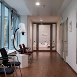
Unser CyberKnife® Zentrum in Soest
Unser CyberKnife® Zentrum finden Sie direkt im Herzen von Nordrhein-Westfalen. Soest ist die Kreisstadt des gleichnamigen Kreises im Regierungsbezirk Arnsberg, direkt zwischen Dortmund und Paderborn. Es ist die erste Krankenhaus-Einrichtung in Deutschland die diese neue Strahlentherapie für Krebspatienten anbieten konnte. Dort begrüßen wir Sie gerne und möchten Ihnen nicht nur eine, sondern die beste Behandlung bieten.
Unsere Partner
Eine sehr gute Vernetzung ist unerlässlich. Wir bedanken uns bei unseren Partnern, die uns diese ermöglichen und stets mit uns, für Sie an einem Strang ziehen.
Einen kleinen Auszug unserer Partner haben wir Ihnen hier aufgeführt.
Strahlentherapie Zentrum Bochum
Klinikum Stadt Soest
Praxis für Strahlentherapie und Radioonkologie Bocholt & Goch
Stereotaxie Zentrum Nordrhein-Westfalen
St. Marien-Hospital Buer
St. Barbara-Klinik Hamm-Heessen
Radiologie Vechta
MVZ Marien-Hospital Wesel
Universitätsklinikum Münster
Klinikum Lippe
Klinikum Herford
Klinikum Hochsauerland
Katholische Hospitalvereinigung Weser-Egge
Universitätsmedizin Essen
MVZ Aurich-Norden GmbH
Onkologisches Centrum Chemnitz
Kontaktieren Sie uns
Nutzen Sie das untenstehende Formular
oder erreichen Sie uns telefonisch.
+49 (0) 2921 66687-0

Indikationen
Rechtliches
Themen
Das Upload-Portal bietet Ihnen die Möglichkeit uns schnell und unkompliziert Ihre Bilddaten (CT, MRT, PET, etc.), Befunde und Berichte zur Verfügung zu stellen.
Kontakt
DEUTSCHES CYBERKNIFE ZENTRUM
Deutsches Zentrum für Stereotaxie
und Präzisionsbestrahlung GmbH
Senator-Schwartz-Ring 8
59494 Soest
Telefon : +49 (0) 2921 66687-0
Telefax: +49 (0) 2921 66687-22
cyberknife@dzsp.de
(für Patienten und Ärzte)
© 2024 Deutsches CyberKnife Zentrum